Accueil / Information / Aller plus loin... / Familles moléculaires / Les polcalcines

Les polcalcines

mercredi 18 août 2010, par Allerdata

Les polcalcines sont des panallergènes polliniques. Ces petites protéines de 6-10 kDa, non glycosylées, font partie d’une famille dénommé 2EF-hand calcium-binding proteins (2EF-CaBP) : elles possèdent, en effet, 2 sites de liaison pour l’ion calcium.

Il existe de nombreuses sortes de protéines liant le calcium, certaines transportant/tamponnant le calcium, d’autres jouant un rôle dans la régulation cellulaire. Les polcalcines s’inscrivent dans une super-famille d’EF-hand, au sein de laquelle on trouve également  :

  • les parvalbumines (poissons, batraciens)
  • les troponines : ex. Tyr p 24 (acarien Tyrophagus), Bla g 6 et Per a 6 (blattes)
  • les protéines S100 : ex. Bos d 3 (bœuf)
  • les sarcoplasmic calcium-binding proteins (SCBP) : dans les Crustacés
  • des allergènes polliniques un peu différents des polcalcines, bien que parfois dénommées aussi polcalcines  :
    • des 3EF-hand, ayant donc 3 sites pour lier le calcium : bouleau (Bet v 3), ambroisie (Amb a 10), armoise
    • des 4EF-hand : olivier (Ole e 8) et Cupressacées (Cup a 4, Jun o 4, Cry j 4, ..)
    • sans être des duplicats des 2EF, les 4EF sont plus apparentées aux polcalcines 2EF que ne le sont les 3EF (cf. cladogramme dans ).

Les polcalcines se différencient d’une autre catégorie de panallergènes des plantes, les profilines : jusqu’à présent on n’a pas identifié de polcalcines IgE-réactives dans d’autres organes végétaux que les pollens.

Mais, comme les profilines, les polcalcines ont une large capacité de réactivité croisée entre elles. De sorte qu’elles constituent bien une catégorie de panallergènes polliniques et que la sensibilisation d’un patient à la polcalcine du pollen X risque fort de générer une réactivité TC, in vitro) vis-à-vis des pollens Y, Z, etc.., même si le patient n’est pas exposé à ces pollens.

Ce qui, bien entendu, va embrouiller le diagnostic, les résultats des tests étant alors volontiers en désaccord avec les données cliniques (ex. période(s) de l’année gênant le patient) .

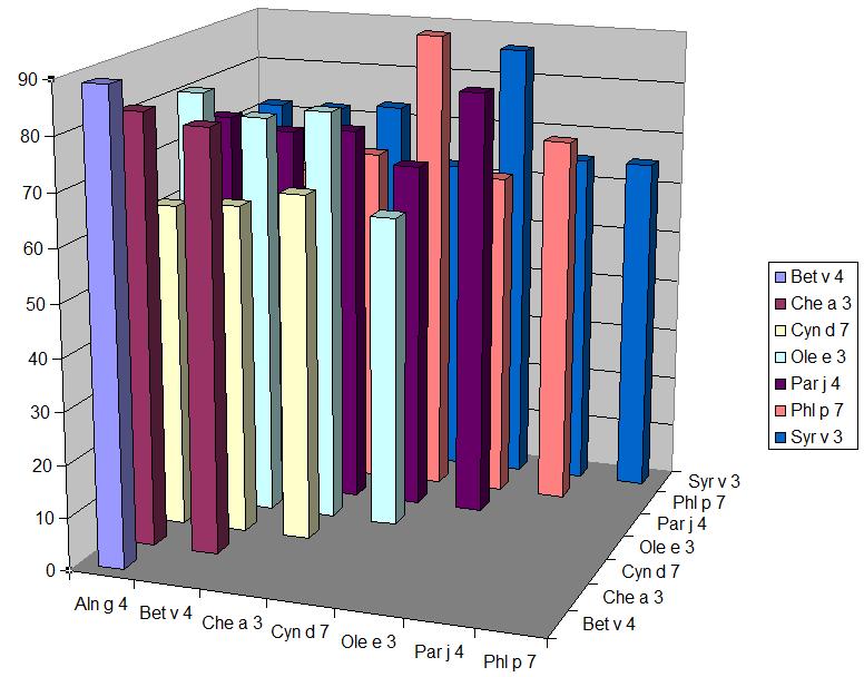
Le tableau ci-dessous montre les % d’identité séquentielle entre polcalcines : ces taux élevés d’homologie sont confirmés au niveau tridimensionnel (épitopes conformationnels) et justifient la large réactivité croisée observée entre ces allergènes etc ..

La réactivité croisée entre polcalcines est-elle qu’il est en pratique suffisant de n’en tester qu’une seule. Par exemple une corrélation élevée (r=0,95) a été trouvée entre les résultants chiffrés (kU/l) de rBet v 4 (bouleau) et de rPhl p 7 (fléole) .

Pour le moment sont testables en CAP rBet v 4 et rPhl p 7. Ces tests sont pertinents pour rechercher une réactivité due à des polcalcines dans la plupart des autres pollens : fagales, graminées, Oléacées, armoise, ambroisie, pariétaire, chénopode, ..

Une polcalcine existe dans le pollen de plantain , mais son IgE-réactivité n’a pas été étudiée. La présence de polcalcines dans les pollens de Cupressacées n’est pas encore établie (NB : ces pollens ont, par contre, des allergènes 4EF-hand).

Les polcalcines (2EF) croisent-elles avec les protéines 3EF ou 4EF ? A priori pas ou peu. Les homologies sont faibles : par exemple les 4 EF comme Cup a 4 (cyprès) ou Ole e 8 (olivier) ont des % d’identité de 48-53% (sur leur portion N-terminale) et 33-34% (sur leur portion C-terminale) avec Phl p 7. Et, bien que peu explorées, les réactivités croisées sont inexistantes , ou dans le sens polcalcine -> 3EF ou 4EF et pas l’autre sens .

Une réactivité vis-à-vis des polcalcines est-elle fréquente ? Le tableau ci-dessous rassemble quelques observations de positivités in vitro :

PaysPollinoseAllergènePositivité %Réf.
Finlande bouleau rBet v 4 5
Suède ^ ^ 8 ^
Autriche 11
Suisse 7
Allemagne boul.+ gram. rBet v 4 6
ou rPhl p 7
France bouleau rBet v 4 6
frêne Fra e 3 3
frêne, etc rChe a 3 24
graminées rPhl p 7 3
Italie bouleau rBet v 4 27
9
graminées rPhl p 7 8
pariétaire Par j 4 5
rPhl p 7 12
ambroisie rAmb a 9 20
et/ou armoise rArt v 5 27
Espagne graminées, etc rPhl p 7 12
olivier Ole e 3 17
graminées rPhl p 7 11
chénopode rChe a 3 46

Comme pour les profilines, les prévalences d’IgE-réactivité pour des polcalcines tendent à croître du Nord au Sud de l’Europe. Avec de fortes variations parfois d’une étude à une autre dans un même pays, ce qui est probablement du à des recrutements de patients assez différents. En effet, la réactivité vis-à-vis des polcalcines semble en grande partie corrélée avec une pluri-réactivité pollinique : une cohorte de patients multi-réactifs pour différents pollens rassemblera une proportion accrue de sujets positifs pour les polcalcines.

Ainsi il n’a été observé aucun cas de positivité pour rBet v 4 ou rPhl p 7 parmi des patients mono-réactifs à une seule catégorie de pollens (fagales ou graminées) .

A l’inverse, la positivité (TC ou in vitro) pour 5 pollens ou plus s’accompagnait d’une réactivité possible pour les polcalcines . La figure ci-dessous, tirée d’un autre travail , présente la répartition des TC cutanés positifs pour la polcalcine (polc) et la profiline (prof) de palmier chez des patients testés avec 9 pollens différents (arbres, graminées, herbacées) :

On pourra noter que les profilines sont un peu plus « rapidement » positives que les polcalcines à mesure que le nombre de pollens positifs augmente.

Il faut souligner cependant qu’une dissociation entre profilines et polcalcine semble fréquente :

  • sur 21 patients positifs in vitro pour une polcalcine et une profiline, seulement 3 étaient simultanément positifs pour les 2 allergènes
  • parmi 16 patients présentant un TC positif pour une polcalcine, 10 étaient négatifs en profiline

So l’on ajoute à cela qu’il est très rare d’observer une mono-positivité pour une polcalcine (ex. rBet v 4 positif avec rBet v 1 et rBet v 2 négatifs), on pourra en déduire la pratique suivante :

  • en cas de pollens positifs peu concordants avec la clinique, on peut tirer parti de la dissociation possible profiline/polcalcine en testant séparément ces 2 allergènes (ex. rPhl p 12 et rPhl p 7 plutôt que le test combiné rPhl p 12+7). On aura ainsi, au besoin, une information supplémentaire en cas de réactions alimentaires associées (rôle d’une profiline ?)
  • il n’est pas pertinent de tester la réactivité aux polcalcines devant une positivité pour 1 ou 2 pollens : cette recherche a toutes chances d’être négative
  • de plus, on ignore la part éventuelle qu’une IgE-réactivité pour une polcalcine pourrait prendre sur le plan clinique dès lors que le patient réagit à des allergènes « majeurs » du pollen (ex. rôle de Bet v 4 chez un patient Bet v 1 positif ??)

L’absence de pertinence clinique d’une positivité pour les polcalcines semble assez bien établie. L’intérêt de cette famille d’allergènes (= plus exactement de protéines IgE-réactives) réside donc dans l’explication de positivités à des pollens qui ne concordent pas avec les données cliniques.

Il n’y a guère que le pollen de chénopode (Chenopodium album, Amaranthacées) qui, selon certains auteurs, recèle une polcalcine (Che a 3) qui, avec la profiline Che a 2, pourrait posséder une pertinence sur le plan clinique . Peut-être est-du à la forme particulière sous laquelle Che a 3 se présente : un tétramère plutôt qu’un dimère (Phl p 7 ) ou un monomère (Bet v 4 ) ?

La recherche d’une IgE-réactivité vis-à-vis d’un panallergène pollinique (profiline, polcalcine) a été suggérée pour guider la décision d’initier ou non une immuno-thérapie : ce point est discuté dans l’article concernant les profilines.

[1] - Wopfner N, Dissertori O, Ferreira F, Lackner P. Calcium-binding proteins and their role in allergic diseases. Immunol Allergy Clin North Am 2007;27:29-44
Calcium-binding proteins (CBPs) are ubiquitous pollen allergens and important food allergens in fish and amphibians. Calcium-binding allergens containing two EF-hands (polcalcins) have been detected and characterized in pollen from trees, grasses, and weeds. Timothy grass Phl p 7 is the most cross-reactive allergen among polcalcins. Although there is cross-reactivity described within the subfamilies of calcium-binding allergens, there are no strong indications for IgE cross-reactivity between CBPs from plants, fish, and humans. Therefore, Phl p 7 could be used as marker to identify multiple pollen-sensitized patients, whereas cod Gad c 1 or carp Cyp c 1 could be selected for the diagnosis of fish allergy. Hom s 4, a calcium-binding autoantigen, might be an interesting candidate to monitor chronic skin inflammation in atopic and nonatopic individuals. Diagnostic tests containing these molecules could allow the identification of most patients sensitized to calcium-binding allergens/antigens. In general, IgE recognition of calcium-binding allergens is influenced by binding or release of calcium ions. This knowledge could be used to engineer hypoallergenic CBPs for specific immunotherapy.
[2] - Tinghino R, Twardosz A, Barletta B, Puggioni EMR, Iacovacci P, Afferni C, et al. Molecular, structural, and immunologic relationships between different families of recombinant calcium-binding pollen allergens. J Allergy Clin Immunol 2002;109:314-320
Background: Calcium-binding plant allergens can be grouped in different families according to the number of calciumbinding domains (EF hands). Objective: We sought to identify pollens containing crossreactive calcium-binding allergens and to investigate structural and immunologic similarities of members belonging to different families of calcium-binding allergens. Methods: By means of multiple sequence alignment and molecular modeling, we searched for structural similarities among pollen allergens with 2 (Phl p 7, timothy grass; Aln g 4, alder), 3 (Bet v 3, birch) and 4 EF hands (Jun o 4, prickly juniper). Purified recombinant Aln g 4 and Jun o 4 were used to determine the prevalence of IgE recognition in 210 patients sensitized to different pollens and to search, by means of ELISA competition, for the presence of cross-reactive epitopes in pollens from 16 unrelated plant species. IgE cross-reactivity among the allergen families was studied with purified rPhl p 7, rAln g 4, rBet v 3, and rJun o 4 and 2 synthetic peptides comprising the N-terminal and C-terminal EF hands of Phl p 7 by means of ELISA competition. Results: Structural similarities were found by using molecular modeling among the allergens with 2, 3, and 4 EF hands. Pollens from 16 unrelated plants contained Aln g 4- and Jun o 4-related epitopes. Twenty-two percent of the patients with multiple pollen sensitization reacted to at least one of the calcium-binding allergens. A hierarchy of IgE cross-reactivity (rPhl p7 > rAln g 4 > rJun o 4 > rBet v 3) could be established that identified rPhl p 7 as the EF-hand allergen containing most IgE epitopes in the population studied. Conclusion: The demonstration that members of different families of calcium-binding plant allergens share similarities suggests that it may be possible to use representative molecules for the diagnosis and therapy of allergies to EF-hand allergens.
[3] - Valenta R, Hayek B, Seiberler S, Bugajska-Schretter A, Niederberger V, Twardosz A, et al. Calcium-binding allergens: from plants to man. Int Arch Allergy Immunol 1998;117:160-166
Calcium-binding proteins contain a variable number of motifs, termed EF-hands, which consist of two perpendicularly placed alpha-helics and an inter-helical loop forming a single calcium-binding site. Due to their ability to bind and transport calcium as well as to interact with a variety of ligands in a calcium-dependent manner, they fulfill important biological functions in eukaryotic cells. After parvalbumin, a three EF-hand fish allergen, calcium-binding allergens were discovered in pollens of trees. grasses and weeds and, recently, as autoallergens in man. Although only a small percentage of atopic individuals displays IgE reactivity to calcium-binding allergens, these allergens may be important because of their ability to cross-sensitize allergic individuals. Confrontation and stability++ as well as IgE recognition of calcium-binding allergens greatly depend on the presence of protein-bound calcium ions. It is thus likely that hypoallergenic derivatives of calcium-binding allergens can be engineered by recombinant DNA technology for immunotherapy++ of sensitized patients. [References: 53]
[4] - Hauser M, Roulias A, Ferreira F, Egger M. Panallergens and their impact on the allergic patient. Allergy Asthma Clin Immunol 2010;6(1):1
ABSTRACT: The panallergen concept encompasses families of related proteins, which are involved in general vital processes and thus, widely distributed throughout nature. Plant panallergens share highly conserved sequence regions, structure, and function. They are responsible for many IgE cross-reactions even between unrelated pollen and plant food allergen sources. Although usually considered as minor allergens, sensitization to panallergens might be problematic as it bears the risk of developing multiple sensitizations. Clinical manifestations seem to be tightly connected with geographical and exposure factors. Future population- and disease-based screenings should provide new insights on panallergens and their contribution to disease manifestations. Such information requires molecule-based diagnostics and will be valuable for developing patient-tailored prophylactic and therapeutic approaches. In this article, we focus on profilins, non-specific lipid transfer proteins, polcalcins, and Bet v 1-related proteins and discuss possible consequences of panallergen sensitization for the allergic patient. Based on their pattern of IgE cross-reactivity, which is reflected by their distribution in the plant kingdom, we propose a novel classification of panallergens into ubiquitously spread "real panallergens" (e.g. profilins) and widespread "eurallergens" (e.g. polcalcins). "Stenallergens" display more limited distribution and cross-reactivity patterns, and "monallergens" are restricted to a single allergen source.
[5] - Radauer C, Breiteneder H. Pollen allergens are restricted to few protein families and show distinct patterns of species distribution. J Allergy Clin Immunol 2006;117:141-147
BACKGROUND: Inhalative allergies are elicited predominantly by pollen of various plant species. However, a classification of the large number of identified pollen allergens is still missing . OBJECTIVE: To analyze pollen allergen sequences with respect to protein family membership, taxonomic distribution of protein families, and interspecies variability . METHODS: Protein family memberships of all plant allergen sequences from the Allergome database were determined by using the Protein Families Database of Alignments and Hidden Markov Models. The taxonomic distribution of pollen allergens was established from the Integrated Taxonomic Information System. Members of abundant pollen allergen families were compared with allergenic and nonallergenic homologues by database similarity searches and multiple sequence alignments . RESULTS: Pollen allergens were classified into 29 of 7868 protein families. Expansins, profilins, and calcium-binding proteins constitute the major pollen allergen families, whereas most plant food allergens belong to the prolamin, cupin, or profilin families. Pollen allergens were revealed to be ubiquitous (eg, profilins), present in certain plant families (eg, pectate lyases), or limited to a single taxon (eg, thaumatin-like proteins). Allergenic plant profilins constitute a highly conserved family with sequence identities of 70% to 85% among each other but low identities of 30% to 40% with nonallergenic profilins from other eukaryotes, including human beings. Similarly, allergenic polcalcins possess sequence identities of 64% to 92% but show low identities of 39% to 42% to related nonallergenic calmodulins and calmodulin-like proteins from vegetative plant tissues and man . CONCLUSION: This classification of pollen allergens into protein families will aid in predicting cross-reactivity, designing comprehensive diagnostic devices, and assessing the allergenic potential of novel proteins.
[6] - Compés E, Quirce S, Villalba M, Hernandez E, Rodriguez R, Sastre J. Sensitization to Robinia pseudoacacia pollen is due to allergenic cross-reactivity with ubiquitous pollen panallergens. EAACI 23th Congress, Amsterdam, 12-16 June, 2004, Poster n°580
Background: Black locust or Robinia pseudoacacia (Rp) is an ornamental tree that belongs to the Leguminosae family. Although it is mainly pollinated by insects, low concentrations of pollen grains from Rp are detected in Madrid during its flowering season. The allergenic composition of this pollen is unknown. Objectives: To study the prevalence of sensitization to Rp pollen in patients with seasonal allergic rhinitis or asthma. To determine the frequency of sensitization to panallergens (profilin, polcalcin, and 1,3-b-glucanase) in patients sensitized to Rp pollen. To investigate if Rp pollen is a primary cause of sensitization or whether it might be implicated through cross-sensitization to other pollens. Methods: Skin prick testing with Rp pollen (ALK-Abelló, Madrid) was performed in 114 consecutive patients with pollinosis. Rp pollen nasal challenge was performed in 10 patients. Specific IgE determinations to Rp pollen, profilin, polcalcin (Che a 3, Ole e 3) and 1,3-b-glucanase (Ole e 9) were performed by ELISA. IgE immunoblotting of Rp pollen and immunoblotting inhibition assays with common pollens were carried out. Results: 64 patients (42.9%) had a positive skin prick test to Rp pollen. Nasal challenges were positive in 5 sensitized patients and negative in 4 controls and in 1 sensitized patient. Specific IgE to Rp pollen was positive in 13 out of 15 (86.6%) subjects with a positive skin prick test. Specific IgE to profilin was found in 9/15 (60%), to polcalcin in 5/15 (33.3%) and to 1,3-b-glucanase in 15/15 (100%) sensitized subjects. Immunoblotting with Rp pollen revealed several IgE binding bands within a wide molecular mass range. Immunoblotting inhibition assays demonstrated that all IgE-binding bands in Rp pollen could be inhibited at some extent by Lolium, Cupressus, Chenopodium, Olea and Rp pollen extracts. Conclusions: We found a high prevalence of Rp pollen sensitization in patients with pollinosis, which is very likely to be due to cross-sensitization to panallergens from other common pollens.
[7] - Verdino P, Barderas R, Villalba M, Westritschnig K, Valenta R, Rodriguez R, et al. Three-dimensional structure of the cross-reactive pollen allergen Che a 3: visualizing cross-reactivity on the molecular surfaces of weed, grass, and tree pollen allergens. J Immunol 2008;180:2313-2321
Two EF-hand calcium-binding allergens (polcalcins) occur in the pollen of a wide variety of unrelated plants as highly cross-reactive allergenic molecules. We report the expression, purification, immunological characterization, and the 1.75-A crystal structure of recombinant Che a 3 (rChe a 3), the polcalcin from the weed Chenopodium album. The three-dimensional structure of rChe a 3 resembles an alpha-helical fold that is essentially identical with that of the two EF-hand allergens from birch pollen, Bet v 4, and timothy grass pollen, Phl p 7. The extensive cross-reactivity between Che a 3 and Phl p 7 is demonstrated by competition experiments with IgE Abs from allergic patients as well as specific Ab probes. Amino acid residues that are conserved for the two EF-hand allergen family were identified in multiple sequence alignments of polcalcins from 15 different plants. Next, the three-dimensional structures of rChe a 3, rPhl p 7, and rBet v 4 were used to identify conserved amino acids with high surface exposition to visualize surface patches as potential targets for the polyclonal IgE Ab response of allergic patients. The essentially identical three-dimensional structures of rChe a 3, rPhl p 7, and rBet v 4 explain the extensive cross-reactivity of allergic patients IgE Abs with two EF-hand allergens from unrelated plants. In addition, analyzing the three-dimensional structures of cross-reactive Ags for conserved and surface exposed amino acids may be a first approach to mapping the conformational epitopes on disease-related Ags that are recognized by polyclonal patient Abs
[8] - Ledesma A, Barderas R, Westritschnig K, Quiralte J, Pascual CY, Valenta R, et al. A comparative analysis of the cross-reactivity in the polcalcin family including Syr v 3, a new member from lilac pollen. Allergy 2006;61:477-484
BACKGROUND: Polcalcins are pollen-specific allergens with two EF-hand calcium-binding sites that exhibit strong cross-reactivity. Our objective was to isolate and express the cDNA coding of the EF-hand calcium-binding allergen from lilac pollen and to study cross-reactivity with other polcalcins from related and nonrelated pollen sources with different specific antibodies and sera from two different populations . METHODS: Specific cDNA was amplified by PCR, cloned and expressed in Escherichia coli. Purification was achieved by gel permeation and ion exchange chromatographies. ELISA titration and inhibition assays were performed using the recombinant forms of Syr v 3, Ole e 3, Che a 3 and Phl p 7 with sera from two Spanish regions with different sensitization profiles, as well as Phl p 7- and Ole e 3-specific polyclonal rabbit antisera, and an Ole e 3-specific monoclonal antibody . RESULTS: Syr v 3 displays two EF-hand consensus sites and 8863 Da of theoretical molecular mass. The allergen consists of 80 residues with identities ranging from 66 to 87% with polcalcins included in this study. Syr v 3, Ole e 3, Che a 3 and Phl p 7 showed a similar IgG- and IgE-binding capacity although differences at quantitative level were observed depending on the population of patients' sera . CONCLUSION: Syr v 3 is a polcalcin with structural and antigenic similarities to the members of this family. Diagnosis of polcalcin-sensitized patients could be performed whatever polcalcin used, whereas for immunotherapy, primary sensitization to a particular allergenic source should be considered.
[9] - Tinghino R, Twardosz A, Barletta B, Puggioni EMR, Iacovacci P, Afferni C, et al. Molecular, structural, and immunologic relationships between different families of recombinant calcium-binding pollen allergens. J Allergy Clin Immunol 2002;109:314-320
Background: Calcium-binding plant allergens can be grouped in different families according to the number of calciumbinding domains (EF hands). Objective: We sought to identify pollens containing crossreactive calcium-binding allergens and to investigate structural and immunologic similarities of members belonging to different families of calcium-binding allergens. Methods: By means of multiple sequence alignment and molecular modeling, we searched for structural similarities among pollen allergens with 2 (Phl p 7, timothy grass; Aln g 4, alder), 3 (Bet v 3, birch) and 4 EF hands (Jun o 4, prickly juniper). Purified recombinant Aln g 4 and Jun o 4 were used to determine the prevalence of IgE recognition in 210 patients sensitized to different pollens and to search, by means of ELISA competition, for the presence of cross-reactive epitopes in pollens from 16 unrelated plant species. IgE cross-reactivity among the allergen families was studied with purified rPhl p 7, rAln g 4, rBet v 3, and rJun o 4 and 2 synthetic peptides comprising the N-terminal and C-terminal EF hands of Phl p 7 by means of ELISA competition. Results: Structural similarities were found by using molecular modeling among the allergens with 2, 3, and 4 EF hands. Pollens from 16 unrelated plants contained Aln g 4- and Jun o 4-related epitopes. Twenty-two percent of the patients with multiple pollen sensitization reacted to at least one of the calcium-binding allergens. A hierarchy of IgE cross-reactivity (rPhl p7 > rAln g 4 > rJun o 4 > rBet v 3) could be established that identified rPhl p 7 as the EF-hand allergen containing most IgE epitopes in the population studied. Conclusion: The demonstration that members of different families of calcium-binding plant allergens share similarities suggests that it may be possible to use representative molecules for the diagnosis and therapy of allergies to EF-hand allergens.
[10] - Egger C, Focke M, Bircher AJ, Scherer K, Mothes-Luksch N, Horak F, et al. The allergen profile of beech and oak pollen. Clin Exp Allergy 2008;38:1688-1696
BACKGROUND: Beech and oak pollen are potential allergen sources with a world-wide distribution . OBJECTIVE: We aimed to characterize the allergen profile of beech and oak pollen and to study cross-reactivities with birch and grass pollen allergens . METHODS: Sera from tree pollen-allergic patients with evidence for beech and oak pollen sensitization from Basel, Switzerland, (n=23) and sera from birch pollen-allergic patients from Vienna, Austria, (n=26) were compared in immunoblot experiments for IgE reactivity to birch (Betula pendula syn. verrucosa), beech (Fagus sylvatica) and oak (Quercus alba) pollen allergens. Subsequently, beech and oak pollen allergens were characterized by IgE inhibition experiments with purified recombinant and natural allergens and with allergen-specific antibody probes. Birch-, beech- and oak pollen-specific IgE levels were determined by ELISA . RESULTS: Beech and oak pollen contain allergens that cross-react with the birch pollen allergens Bet v 1, Bet v 2 and Bet v 4 and with the berberine bridge enzyme-like allergen Phl p 4 from timothy grass pollen. Sera from Swiss and Austrian patients exhibited similar IgE reactivity profiles to birch, beech and oak pollen extracts. IgE levels to beech and oak pollen allergens were lower than those to birch pollen allergens . CONCLUSION: IgE reactivity to beech pollen is mainly due to cross-reactivity with birch pollen allergens, and a Phl p 4-like molecule represented another predominant IgE-reactive structure in oak pollen. The characterization of beech and oak pollen allergens and their cross-reactivity is important for the diagnosis and treatment of beech and oak pollen allergy.
[12] - Hayek B, Vangelista L, Pastore A, Sperr WR, Valent P, Vrtala S, et al. Molecular and immunologic characterization of a highly cross-reactive two EF-hand calcium-binding alder pollen allergen, Aln g 4: structural basis for calcium-modulated IgE recognition. J Immunol 1998;161:7031-7039
Serum IgE was used to isolate a cDNA coding for a 9.4-kDa two EF-hand calcium-binding allergen, Aln g 4, from a lambda gt11 expression cDNA library constructed from alder (Alnus glutinosa) pollen. rAln g 4 was overexpressed in Escherichia coli and purified to homogeneity. It reacted with serum IgE from 18% of pollen-allergic patients (n = 122); shared IgE epitopes with homologous allergens present in tree, grass, and weed pollens; and thus belongs to a family of highly cross-reactive pollen allergens. Exposure of two E. coli-expressed rAln g 4 fragments comprising amino acids 1-41 and 42-85 to patients' IgE Abs, as well as to a rabbit antiserum raised against purified rAln g 4, indicated that most of the B cell epitopes reside in the N-terminal portion of the protein. IgE recognition of Aln g 4 was strongly modulated by the presence or absence of calcium. Circular dichroism analysis of rAln g 4 revealed that the protein consisted mostly of alpha helical secondary structure and possessed a remarkable thermal stability and refolding capacity, a property that was greatly reduced after calcium depletion. Circular dichroism analysis of the calcium-bound and apo form of rAln g 4 indicated that calcium-induced modulation of IgE binding could be due to changes in the protein conformation. Purified rAln g 4 elicited dose-dependent basophil histamine release and immediate type skin reactions in sensitized patients. It may hence be useful for allergy diagnosis and for specific immunotherapy.
[13] - Barderas R, Villalba M, Pascual CY, Batenero E, Rodriguez E. Profilin (Che a 2) and polcalcin (Che a 3) are relevant allergens of Chenopodium album pollen: Isolation, amino acid sequences, and immunologic properties. J Allergy Clin Immunol 2004;113:1192-1198
Background Little is known about the molecular properties of chenopod allergens. Recently, profilin and 2 EF-hand calcium-binding protein (polcalcin) have been shown to play a role in chenopod pollinosis. Objective : We sought to analyze these panallergens in chenopod pollen and to evaluate their involvement in the allergy to this biologic source. Method s : Profilin and polcalcin were purified to homogeneity and characterized by using spectrometric and chemical methods. Immunologic analyses were performed by means of immunoblotting, ELISA, and competitive inhibition assays with olive profilinˆ and polcalcin-specific rabbit polyclonal antibodies and sera from patients with chenopod allergy. cDNAs encoding these proteins were cloned by means of PCR and sequenced. Result s : Purified Che a 2 (profilin) and Che a 3 (polcalcin) exhibited prevalences of 55% and 46%, respectively, in patients (n=104) hypersensitive to chenopod pollen. Both purified allergens individually inhibited the IgE binding to the whole pollen extract and showed strong cross-reactivity with the corresponding olive pollen profilin (Ole e 2) and polcalcin (Ole e 3). Chenopod profilin consists of a 131-amino-acid chain that displays identities of approximately 75% and 82% with pollen and food profilins, respectively. Che a 3 (86 amino acids) displays similarity (65% to 82% identity) with polcalcins from pollens of olive, birch, alder, rapeseed, and timothy. Conclusion : Profilin and polcalcin are relevant panallergens in chenopod pollen and good candidates to be involved in IgE cross-reactivity with other pollen sources, thus explaining the highly frequent polysensitization of patients allergic to chenopod.
[14] - Rossi RE, Monasterolo G, Monasterolo S. Measurement of IgE antibodies against purified grass-pollen allergens (Phl p 1, 2, 3, 4, 5, 6, 7, 11, and 12) in sera of patients allergic to grass pollen. Allergy 2001;56:1180-1185
Background: Current allergy diagnosis is performed with allergen extracts which contain a variety of allergenic and nonallergenic components. The availability of highly purified and well-characterized allergen molecules seems to be an advantage of component-based diagnosis. Methods: With the immunoenzymatic CAP FEIA System, we measured specific IgE levels to the recombinant allergens rPhl p 1, rPhl p 2, rPhl p 5, rPhl p 6, rPhl p 7, rPhl p 11, rPhl p 12, and native Phl p 4 in 77 sera of patients allergic to grass pollen, in order to evaluate the IgE-binding frequency to these purified grass-pollen allergens and their relationship to rBet v 4, rBet v 2, and other allergens. Results: The frequency of sensitization was as follows: rPhl p 1=93.5%; rPhl p 2=67.5%; rPhl p 5=72.7%; rPhl p 6=68.8%; rPhl p 7=7.8%; rPhl p 11=53.2%; rPhl p 12=35.1%; and native Phl p 4=88.3%. As expected, rPhl p 7 and rPhl p 12 had a very good correlation (Spearman's r) with Bet v 4 (r=0.95%, P<0.05) and rBet v 2 (r=0.99, P<0.05), respectively. Good correlations of rPhl p 12 with papain (r=0.93, P<0.05), latex (r=0.92, P<0.05), and bromelain (r=0.86, P<0.05) were found. Highly variable individual sensitization patterns were observed. Conclusions: A new clinical approach has allowed the determination of specific allergograms for the different patients and may therefore be of great importance for more specific diagnosis. The use of component-resolved diagnostics may be useful to evaluate the allergen content of an extract for immunotherapy by monitoring patient's IgE and IgG directed to relevant allergens.
[15] - Grote M, Westritschnig K, Valenta R. Immunogold Electron Microscopic Localization of the 2 EF-Hand Calcium-Binding Pollen Allergen Phl p 7 and its Homologues in Pollens of Grasses, Weeds and Trees. Int Arch Allergy Immunol 2008;146:113-121
BACKGROUND: The 2 EF-hand calcium-binding allergen from timothy grass pollen, Phl p 7, contains the majority of relevant IgE epitopes among calcium-binding allergens occurring in pollen species of different plants . OBJECTIVE: To describe the ultrastructural localization of Phl p 7 allergen in timothy grass pollen and its homologues in a broad spectrum of allergologically relevant pollens from grasses (timothy grass, rye grass), trees (birch, alder, olive) and weeds (mugwort, ribwort, ragweed) commonly growing in Europe. MATERIALS AND METHODS: Mature pollens from 8 different plant species were collected and anhydrously prepared for transmission electron microscopy. In ultrathin sections, allergens were localized using an antibody prepared against a Phl p 7-derived peptide comprising the C-terminal half of the Phl p 7 wild-type molecule in combination with a secondary antibody coupled to 10-nm colloidal gold particles . RESULTS: Phl p 7 and Phl p 7 homologues were detected in pollen from each of the 8 pollen species investigated. The allergens were found in the cytoplasm of the pollen grains (cytoplasmic matrix, mitochondria, nuclei) and in the pollen wall (preferably the exine). Reserve materials were unlabeled . CONCLUSIONS: The 2 EF-hand calcium-binding allergen Phl p 7 from timothy grass and its homologues can be localized in all pollen species under investigation. This finding confirms that Phl p 7 is a marker allergen for sensitization of patients to a novel family of 2 EF-hand calcium-binding pollen allergens occurring in a number of important allergenic plants in Europe.
[16] - Wopfner N, Gruber P, Wallner M, Briza P, Ebner C, Mari A, et al. Molecular and immunological characterization of novel weed pollen pan-allergens. Allergy 2008;63:872-881
BACKGROUND: Pan-allergens like profilins, calcium-binding proteins (CBPs), and nonspecific lipid transfer proteins have been suggested as possible specific markers for multiple pollen sensitizations, and could be used to predict cross-sensitization/poly-sensitization to several pollen allergens. Therefore, the purification and characterization of cross-reacting allergens in pollen is an extremely important task towards correct allergy diagnosis . METHODS: New pan-allergens were identified by screening a ragweed pollen cDNA library with sera of patients allergic to mugwort pollen. Resulting proteins were cloned, expressed, purified and characterized . RESULTS: We report complete cDNA sequences of two profilin isoforms (Amb a 8.01 and Amb a 8.02), two isoforms of a 2EF-hand CBP (Amb a 9.01 and Amb a 9.02), a new 3EF-hand CBP (Amb a 10) from ragweed pollen and a 2EF-hand CBP from mugwort (Art v 5). All these proteins were expressed in Escherichia coli, purified to homogeneity and characterized by biochemical and immunological means . CONCLUSIONS: The identified proteins are novel pan-allergens and can be used as diagnostic markers for polysensitization and used in component-resolved diagnosis.
[17] - Smith PM, Xu H, Swoboda I, Singh MB. Identification of a Ca2+ binding protein as a new Bermuda grass pollen allergen Cyn d7: IgE cross-reactivity with oilseed rape pollen allergen Bra r1. Int Arch Allergy Immunol 1997;114:265-271
cDNA clones encoding two isoforms of an allergen from pollen of Bermuda grass (Cynodon dactylon) have been isolated using IgE from allergic patients. Homologous transcripts are present in pollen of 15 other grasses tested. This allergen, tentatively designated as Cyn d 7, contains two calcium binding domains and shows significant sequence similarity with other Ca2+ binding pollen allergens, namely Bet v 4 from birch and Bra r 1 from oilseed rape. Approximately 10% of allergic sera tested showed IgE reactivity to this allergen. IgE cross-reactivity was observed between this allergen and Bra r 1 of oilseed rape. IgE reactivity of this allergen requires protein-bound Ca2+. Using IgE affinity-purified from the recombinant allergen to probe Western blots of pollen extracts Cyn d 7 has been identified as a 12 kDA protein.
[18] - Ledesma A, González E, Pascual CY, Quiralte J, Villalba M, Rodríguez R. Are Ca2+-binding motifs involved in the immunoglobin E-binding of allergens ? Olive pollen allergens as model of study. Clin Exp Allergy 2002;32:1476-1483
BackgroundSeveral Ca2+-binding proteins, which possess EF-hand sites with a high sequence similarity, have been found to be able to induce Type-I allergy. ObjectiveTo study whether the common EF-hand sequential motifs can be involved in the IgE-reactivity of these proteins, thus being responsible of a degree of cross-reactivity among different Ca2+-binding proteins. MethodsTwo olive pollen allergens, Ole e 3 and Ole e 8, have been used in the study. Parvalbumin and calmodulin were included in immunological analyses. Sera from patients allergic to olive pollen, as well as Ole e 3- and Ole e 8-specific rabbit antisera were used in indirect enzyme-linked immunosorbent assay (ELISA), ELISA inhibition assays and immunobloting. Conformational analyses (circular dichroism spectra and thermal stability) and specific immunodetection assays were performed in the presence and the absence of Ca2+. Chemical breakdown and high-performance liquid chromatography (HPLC) was used to obtain fragments from Ole e 3 containing a single EF-hand motif. ResultsThirty-four (17%) and 16 (8.2%) out of 195 sera from patients allergic to olive pollen contained specific IgE against Ole e 3 and Ole e 8, respectively. The IgE-binding of 12 allergic sera diminished up to 22% for Ole e 3 and to 82% for Ole e 8, when depleted Ca2+. A pool of these sera recognized the two olive allergens and parvalbumin, but at very different extent. Inhibition of the IgE-binding was only achieved between two olive allergens. No structural relationships between Ole e 3 and Ole e 8 were established when specific polyclonal antisera against both proteins were used. ConclusionEF-hand Ca2+-binding sites can not be considered as general allergenic motifs responsible for the cross-reactivity between Ca2+-binding allergens. Different families of Ca2+-binding allergens have specific epitopes that could be involved in the cross-reactivity among members of the same family.
[19] - Tinghino R, Twardosz A, Barletta B, Puggioni EMR, Iacovacci P, Afferni C, et al. Molecular, structural, and immunologic relationships between different families of recombinant calcium-binding pollen allergens. J Allergy Clin Immunol 2002;109:314-320
Background: Calcium-binding plant allergens can be grouped in different families according to the number of calciumbinding domains (EF hands). Objective: We sought to identify pollens containing crossreactive calcium-binding allergens and to investigate structural and immunologic similarities of members belonging to different families of calcium-binding allergens. Methods: By means of multiple sequence alignment and molecular modeling, we searched for structural similarities among pollen allergens with 2 (Phl p 7, timothy grass; Aln g 4, alder), 3 (Bet v 3, birch) and 4 EF hands (Jun o 4, prickly juniper). Purified recombinant Aln g 4 and Jun o 4 were used to determine the prevalence of IgE recognition in 210 patients sensitized to different pollens and to search, by means of ELISA competition, for the presence of cross-reactive epitopes in pollens from 16 unrelated plant species. IgE cross-reactivity among the allergen families was studied with purified rPhl p 7, rAln g 4, rBet v 3, and rJun o 4 and 2 synthetic peptides comprising the N-terminal and C-terminal EF hands of Phl p 7 by means of ELISA competition. Results: Structural similarities were found by using molecular modeling among the allergens with 2, 3, and 4 EF hands. Pollens from 16 unrelated plants contained Aln g 4- and Jun o 4-related epitopes. Twenty-two percent of the patients with multiple pollen sensitization reacted to at least one of the calcium-binding allergens. A hierarchy of IgE cross-reactivity (rPhl p7 > rAln g 4 > rJun o 4 > rBet v 3) could be established that identified rPhl p 7 as the EF-hand allergen containing most IgE epitopes in the population studied. Conclusion: The demonstration that members of different families of calcium-binding plant allergens share similarities suggests that it may be possible to use representative molecules for the diagnosis and therapy of allergies to EF-hand allergens.
[20] - Movérare R, Westritschnig K, Svensson M, Hayek B, Bende M, Pauli G, et al. Different IgE Reactivity Profiles in Birch Pollen-Sensitive Patients from Six European Populations Revealed by Recombinant Allergens: An Imprint of Local Sensitization. Int Arch Allergy Immunol 2002;128:325-335
Background: Sensitivity to birch pollen allergens is a common feature among European patients with seasonal pollen allergy. In this in vitro study, we examined the specific serum IgE binding profiles to individual birch pollen allergens in birch-sensitive patients from six European populations. Methods: The study included 242 patients from Finland, Sweden, Austria, France, Switzerland and Italy. All suffered from seasonal rhinoconjunctivitis and/or asthma. Their sera were analyzed for specific IgE reactivity to individual birch pollen allergens (recombinant Bet v 1, Bet v 2 and Bet v 4) and natural birch pollen extract using Pharmacia CAP System and immunoblotting. Results: Almost all Finnish, Swedish and Austrian sera contained IgE specific for Bet v 1 (98%). Bet v 1-specific IgE antibodies were found in 90% of the French sera, and in 65 and 62% of the sera from Switzerland and Italy, respectively. Few Finnish (2%) and Swedish (12%) patients had IgE to Bet v 2, while Bet v 2 reactivity was more common in the other populations (20-43%). Reactivity to Bet v 4 was rare in all populations (5-11%) except for the Italian patients, in whom 3 of 11 sera were positive (27%). The immunoblot results supported the specific IgE profiles obtained with Pharmacia CAP System showing a broader IgE reactivity profile in patients from central and southern Europe as compared to northern Europe. Conclusion: Component-resolved allergy diagnosis with recombinant allergens reveals that the IgE reactivity profiles to individual birch pollen allergens vary between European populations. This observation may be explained by sensitization to different allergen sources and will have an impact on allergen-specific prevention and therapy strategies.
[21] - Schierz J, Burow G. Determination of antibody pattern with recombinant allergen components in pollen allergic patients. Allergy Clin Immunol Int 2005;17(Suppl. 1):205
Quantitative determination of specific IgE antibodies to recombinant allergen components allows to establish the individual sensitization profiles of allergic patients. The presence of IgE antibodies to crossreactive components can be used to predict sensitization to related allergen sources. The aim of the study is to determine the IgE antibody pattern in sera of grass and birch pollen allergic patients. Methods: Serum samples from 151 patients were analyzed for IgE antibodies to recombinant major allergens of timothy and birch, Phl p1, Phl p5, Bet v1, the profilins Phl p12, Bet v2, and the calcium-binding proteins Phl p7 and Bet v4 with UniCAP 100 System and the recombinant ImmunoCAP allergens (Pharmacia Diagnostics, Freiburg, Germany). Serum samples with levels >5kU/l to timothy and/or birch pollen were selected. Results: Serum samples of 44 patients were positive for the major allergens of both timothy and birch pollen, Phl p1, Phl p5, Bet v1, and negative for Phl p7, Phl p12, Bet v2 and Bet v4. 51 samples were only positive for Phl p1 and Phl p5, negative for birch, 30 samples were only positive for Bet v1, negative for timothy. 6 samples positive for timothy, levels between 5.7-9.4 kU/l were only positive for the additionally tested component Phl p4. One sample positive for timothy and rye pollen was only positive for Phl p7 and Phl p12, but negative for the major components. In 19 samples IgE antibodies to both timothy and birch pollen major allergens but also to profilins and calcium-binding proteins could be detected, partly with lower levels for the major allergen components. Conclusion: In 125 of 151 samples IgE antibodies were found to only the major allergen components of timothy and/or birch pollen. In 26 samples antibodies to profilins and/or calcium-binding proteins were detected, partly with negative or lower levels for the major allergens. The measurement of IgE antibodies with recombinant allergen components is useful for the detection of individual sensitization pattern to predict crossreactivity and might be of importance for the selection of the patients for specific immunotherapy.
[22] - Movérare R, Westritschnig K, Svensson M, Hayek B, Bende M, Pauli G, et al. Different IgE Reactivity Profiles in Birch Pollen-Sensitive Patients from Six European Populations Revealed by Recombinant Allergens: An Imprint of Local Sensitization. Int Arch Allergy Immunol 2002;128:325-335
Background: Sensitivity to birch pollen allergens is a common feature among European patients with seasonal pollen allergy. In this in vitro study, we examined the specific serum IgE binding profiles to individual birch pollen allergens in birch-sensitive patients from six European populations. Methods: The study included 242 patients from Finland, Sweden, Austria, France, Switzerland and Italy. All suffered from seasonal rhinoconjunctivitis and/or asthma. Their sera were analyzed for specific IgE reactivity to individual birch pollen allergens (recombinant Bet v 1, Bet v 2 and Bet v 4) and natural birch pollen extract using Pharmacia CAP System and immunoblotting. Results: Almost all Finnish, Swedish and Austrian sera contained IgE specific for Bet v 1 (98%). Bet v 1-specific IgE antibodies were found in 90% of the French sera, and in 65 and 62% of the sera from Switzerland and Italy, respectively. Few Finnish (2%) and Swedish (12%) patients had IgE to Bet v 2, while Bet v 2 reactivity was more common in the other populations (20-43%). Reactivity to Bet v 4 was rare in all populations (5-11%) except for the Italian patients, in whom 3 of 11 sera were positive (27%). The immunoblot results supported the specific IgE profiles obtained with Pharmacia CAP System showing a broader IgE reactivity profile in patients from central and southern Europe as compared to northern Europe. Conclusion: Component-resolved allergy diagnosis with recombinant allergens reveals that the IgE reactivity profiles to individual birch pollen allergens vary between European populations. This observation may be explained by sensitization to different allergen sources and will have an impact on allergen-specific prevention and therapy strategies.
[23] - Poncet P, Sénéchal H, Clément G, Purohit A, Sutra JP, Desvaux FX et al. Evaluation of ash pollen sensitization pattern using proteomic approach with individual sera from allergic patients. Allergy 2010;65:571-580
Food allergies, defined as an adverse immune response to food proteins, affect as many as 6% of young children and 3%-4% of adults in westernized countries, and their prevalence appears to be rising. In addition to well-recognized acute allergic reactions and anaphylaxis triggered by IgE antibody-mediated immune responses to food proteins, there is an increasing recognition of cell-mediated disorders such as eosinophilic gastroenteropathies and food protein-induced enterocolitis syndrome. We are gaining an increasing understanding of the pathophysiology of food allergic disorders and are beginning to comprehend how these result from a failure to establish or maintain normal oral tolerance. Many food allergens have been characterized at a molecular level, and this knowledge, combined with an increasing appreciation of the nature of humoral and cellular immune responses resulting in allergy or tolerance, is leading to novel therapeutic approaches. Currently, management of food allergies consists of educating the patient to avoid ingesting the responsible allergen and initiating therapy if ingestion occurs. However, numerous strategies for definitive treatment are being studied, including sublingual/oral immunotherapy, injection of anti-IgE antibodies, cytokine/anticytokine therapies, Chinese herbal therapies, and novel immunotherapies utilizing engineered proteins and strategic immunomodulators.
[26] - Movérare R, Westritschnig K, Svensson M, Hayek B, Bende M, Pauli G, et al. Different IgE Reactivity Profiles in Birch Pollen-Sensitive Patients from Six European Populations Revealed by Recombinant Allergens: An Imprint of Local Sensitization. Int Arch Allergy Immunol 2002;128:325-335
Background: Sensitivity to birch pollen allergens is a common feature among European patients with seasonal pollen allergy. In this in vitro study, we examined the specific serum IgE binding profiles to individual birch pollen allergens in birch-sensitive patients from six European populations. Methods: The study included 242 patients from Finland, Sweden, Austria, France, Switzerland and Italy. All suffered from seasonal rhinoconjunctivitis and/or asthma. Their sera were analyzed for specific IgE reactivity to individual birch pollen allergens (recombinant Bet v 1, Bet v 2 and Bet v 4) and natural birch pollen extract using Pharmacia CAP System and immunoblotting. Results: Almost all Finnish, Swedish and Austrian sera contained IgE specific for Bet v 1 (98%). Bet v 1-specific IgE antibodies were found in 90% of the French sera, and in 65 and 62% of the sera from Switzerland and Italy, respectively. Few Finnish (2%) and Swedish (12%) patients had IgE to Bet v 2, while Bet v 2 reactivity was more common in the other populations (20-43%). Reactivity to Bet v 4 was rare in all populations (5-11%) except for the Italian patients, in whom 3 of 11 sera were positive (27%). The immunoblot results supported the specific IgE profiles obtained with Pharmacia CAP System showing a broader IgE reactivity profile in patients from central and southern Europe as compared to northern Europe. Conclusion: Component-resolved allergy diagnosis with recombinant allergens reveals that the IgE reactivity profiles to individual birch pollen allergens vary between European populations. This observation may be explained by sensitization to different allergen sources and will have an impact on allergen-specific prevention and therapy strategies.
[27] - Rossi RE, Monasterolo G, Monasterolo S. Detection of specific IgE antibodies in the sera of patients allergic to birch pollen using recombinant allergens Bet v 1, Bet v 2, Bet v 4: evaluation of different IgE reactivity profiles. Allergy 2003;58:929-932
BACKGROUND: Birch pollen is a significant cause of immediate hypersensitivity among susceptible subjects in temperate climates, affecting 5-54% of the population in western Europe. We examined the specific serum IgE antibodies towards recombinant allergens Bet v 1, Bet v 2 and Bet v 4 in birch-sensitive patients from the province of Cuneo, north-west Italy . METHODS: Sera were obtained from 372 patients with symptomatic birch pollen-induced allergic rhinitis and/or asthma. A subgroup of these patients suffered from oral allergy syndrome after eating apple. Their sera were evaluated for specific IgE against natural birch pollen and apple extract, as well as Bet v 1, Bet v 2 and Bet v 4 using Pharmacia CAP system (Pharmacia, Uppsala, Sweden) . RESULTS: Of 372 patients 215 (57.80%) had serum-specific IgE towards Bet v 1. A total of 166 sera (44.62%) contained serum-specific IgE to Bet v 2, while Bet v 4 IgE reactivity was documented in 35 subjects (9.41%). Moreover, 146 (39.25%) patients were monosensitized to Bet v 1; 96 (25.81%) patients were monosensitized to Bet v 2; only four sera (1.08%) contained specific IgE towards Bet v 4. Thirty-nine sera (11.02%) did not contain specific IgE to these individual birch pollen allergens. Of course, all 372 sera (100%) had specific IgE against natural birch pollen extract, of which 162 (43.55%) contained specific IgE to apple extract (75.35% of Bet v 1 positive sera) . CONCLUSION: In this study we observed that three birch pollen recombinant allergens alone, could sufficiently identify 90% of birch pollen-sensitive patients. Therefore, for a more precise IgE profile of patients allergic to birch, further purified birch pollen allergens (i.e. Bet v 6, Bet v 7, Bet v 8) will be required.
[28] - Rossi RE, Monasterolo G, Monasterolo S. Measurement of IgE antibodies against purified grass-pollen allergens (Phl p 1, 2, 3, 4, 5, 6, 7, 11, and 12) in sera of patients allergic to grass pollen. Allergy 2001;56:1180-1185
Background: Current allergy diagnosis is performed with allergen extracts which contain a variety of allergenic and nonallergenic components. The availability of highly purified and well-characterized allergen molecules seems to be an advantage of component-based diagnosis. Methods: With the immunoenzymatic CAP FEIA System, we measured specific IgE levels to the recombinant allergens rPhl p 1, rPhl p 2, rPhl p 5, rPhl p 6, rPhl p 7, rPhl p 11, rPhl p 12, and native Phl p 4 in 77 sera of patients allergic to grass pollen, in order to evaluate the IgE-binding frequency to these purified grass-pollen allergens and their relationship to rBet v 4, rBet v 2, and other allergens. Results: The frequency of sensitization was as follows: rPhl p 1=93.5%; rPhl p 2=67.5%; rPhl p 5=72.7%; rPhl p 6=68.8%; rPhl p 7=7.8%; rPhl p 11=53.2%; rPhl p 12=35.1%; and native Phl p 4=88.3%. As expected, rPhl p 7 and rPhl p 12 had a very good correlation (Spearman's r) with Bet v 4 (r=0.95%, P<0.05) and rBet v 2 (r=0.99, P<0.05), respectively. Good correlations of rPhl p 12 with papain (r=0.93, P<0.05), latex (r=0.92, P<0.05), and bromelain (r=0.86, P<0.05) were found. Highly variable individual sensitization patterns were observed. Conclusions: A new clinical approach has allowed the determination of specific allergograms for the different patients and may therefore be of great importance for more specific diagnosis. The use of component-resolved diagnostics may be useful to evaluate the allergen content of an extract for immunotherapy by monitoring patient's IgE and IgG directed to relevant allergens.
[29] - Bonura A, Gulino L, Trapani A, Di Felice G, Tinghino R, Amoroso S, et al. Isolation, expression and immunological characterization of a calcium-binding protein from Parietaria pollen. Mol Immunol 2008;45:2465-2473
The diagnosis and therapy of allergic disorders are usually performed with crude extracts which are a heterogeneous mixture of proteins with different allergenic potency. The knowledge of the allergenic composition is a key step for diagnostic and therapeutic options. Parietaria judaica pollen represents one of the main sources of allergens in the Mediterranean area and its major allergens have already been identified (Par j 1 and Par j 2). In addition, inhibition studies performed using a calcium-binding protein (CBP) from grass pollen (Phl p 7) showed the presence of a homologue of this cross-reactive allergen in the Parietaria extract. Screening of a cDNA library allowed us to isolate a 480bp cDNA containing the information for an 87 AA long protein with high level of homology to calcium-binding proteins from other allergenic sources. It was expressed as a recombinant allergen in Escherichia coli and purified by affinity chromatography. Its expression allowed us to study the prevalence of this allergen in a population of allergic patients in southern Europe. Immunoblotting and inhibition studies showed that this allergen shares a pattern of IgE epitopes in common with other 2-EF-hand calcium-binding proteins from botanically non-related species. The immunological properties of the Pj CBP were investigated by CD63 activation assay and CFDA-SE staining. In conclusion, DNA recombinant technology allowed the isolation, expression and immunological characterization of a cross-reactive calcium-binding protein allergen from Parietaria judaica pollen.
[30] - Rossi RE, Monasterolo G, Coco G, Operti D. Possible Relationship between Systemic Side Effects and Sensitization to rPar j 2 in Allergic Patients Submitted to an Ultra-Rush (20 min) Sublingual Immunotherapy and Selected by Component Resolved Diagnosis. Int Arch Allergy Immunol 2005;138:105-110
BACKGROUND: The pollen of Parietaria spp., a weed of the Urticaceae family, is a major cause of respiratory allergy in the Mediterranean area, where the most common species are Parietaria judaica and Parietaria officinalis. In this study, we evaluated the specific serum IgE-binding profiles to individual P. judaica pollen recombinant major allergen, and Phleum pratense cytoskeletal profilin and a 2-EF-hand calcium-binding allergen homologous to cross-reactive Parietaria pollen allergens, in patients allergic to pollen with positive skin test towards Parietaria spp. extract . METHODS: The present observation included 220 patients from the province of Cuneo, north-west Italy, all suffering from rhino-conjunctivitis and/or asthma selected on the basis of skin test positive to P. judaica extract. The sera were evaluated for specific IgE reactivity to P. judaica pollen major recombinant(r) allergen Par j 2, Phleum pratense pollen allergens rPhl p 7 (2-EF-hand calcium binding protein) and rPhl p 12 (profilin), both identified as cross-reactive Parietaria spp. allergens, using Pharmacia CAP System. Out of 220 patients, 37 patients with IgE reactivity to rPar j 2 and 105 patients sensitized to at least one timothy pollen major allergen (i. e. rPhl p 1, rPhl p 2, natural Phl p 4 and rPhl p 6) were submitted to an ultra-rush protocol of sublingual immunotherapy (SLIT). The occurrence of adverse reactions were evaluated in both groups . RESULTS: All 220 patients with pollinosis and positive in vivo skin prick tests had in vitro positive CAP results to P. judaica natural extract. On the contrary, in these patients the prevalence of Par j 2-specific IgE was only 33.2% (73/220). In fact, 116/220 (52.7%) patients with serum specific IgE to crude Parietaria pollen extract had specific IgE to Phl p 12, 18/220 (8.1%) subjects with specific IgE to rPhl p 12 also exhibited specific IgE to Phl p 7 and 26/220 (11.8%) subjects had specific IgE against rPhl p 7. Particularly, geometric mean (25th-75th percentile) of specific IgE to rPar j 2 were as follows: 2.87 kUA/l (1.005-7.465). Out of 73 patients with specific IgE to rPar j 2, 7 subjects (9.6%) had also specific IgE to rPhl p 7, 12 (16.4%) had specific IgE to rPhl p 12 and 4 (4.1%) patients had specific IgE to both recombinant allergens. Of 37 patients under an ultra-rush protocol of SLIT, 3 subjects (8.1%) experienced generalized urticaria, and 1 of them also had diarrhea 3 h after the last dose of Parietaria judaica extract oral-vaccine administration. On the contrary, no systemic reactions were observed in 105 patients after Phleum pratense extract oral intake after a similar ultra-rush SLIT protocol (p = 0.0046) . CONCLUSIONS: In the light of present findings, allergen extract-based diagnosis, in vivo and in vitro, cannot discriminate allergic patients that are genuinely sensitized to Parietaria spp. major allergens or to other major allergens to which current immunotherapeutic allergy extracts are standardized. Therefore, in vitro component resolved diagnosis is the unique tool to define the disease eliciting molecule(s). Finally, during sublingual immunotherapy, not only the dose of allergen, but also the biochemical characteristic of the major allergen administered may be an important factor in determining possible systemic reactions.
[31] - Asero R, Wopfner N, Gruber P, Gadermaier G, Ferreira F. Artemisia and Ambrosia hypersensitivity: co-sensitization or co-recognition ? Clin Exp Allergy 2006;36:658-665
BACKGROUND: Ragweed and mugwort have nearly identical flowering periods. Clinical and serological studies showed that ragweed and mugwort sensitization are often associated and this poses relevant clinical problems in patients for whom specific immunotherapy is warranted . OBJECTIVE: To establish whether the concurrent ragweed and mugwort pollen hypersensitivity is the result of co-sensitization or of co-recognition by using purified recombinant allergens . METHODS: Sensitization to ragweed and mugwort pollen was assessed by skin prick test (SPT) in all patients reporting allergic symptoms in August and September. IgE reactivity of sera from 42 patients (26 Amb+/Art+, 14 Amb+/Art-, and two Amb-/Art+) to ragweed and mugwort pollen extract as well as to several recombinant ragweed (rAmb a 1, rAmb a 5, rAmb a 6, rAmb a 8, rAmb a 9, and Amb a 10) and mugwort (rArt v 1, rArt v 4, rArt v 5, rArt v 6, and three EF-hand calcium-binding protein) allergens was detected by dot-blot and ELISA analyses . RESULTS: IgE reactivity of 372 weed pollen-allergic patients was studied. Mugwort reactivity was strongly associated with ragweed hypersensitivity: only 10/147 (7%) mugwort-hypersensitive patients were not sensitized to ragweed, whereas 225/362 (62%) ragweed-hypersensitive patients were not sensitized to mugwort. In vitro, 90% of ragweed-allergic patients reacted with rAmb a 1. Reactivity to other ragweed allergens ranged between 20% and 35%. Forty-six percent of the mugwort-sensitized patients recognized rArt v 1%, 25% reacted to Art v 4, Art v 5, and Art v 6, and 7% recognized the three-EF hand calcium-binding protein. Immunoblot inhibition experiments showed that pre-incubation with ragweed pollen extract only weakly decreased IgE reactivity to mugwort allergens . CONCLUSION: Patients showing both ragweed- and mugwort-positive SPT and/or RAST are co-sensitized. Future studies will establish whether IgE reactivity translates into clinical symptoms and, hence, if co-sensitized patients should undergo specific immunotherapy with extracts of both mugwort and ragweed pollen.
[32] - Luengo O, Mollá R, Gámez C, Cardona V, López E, Sastre B, et al. Allergenicity and cross-reactivity of Senecio pollen: identification of novel allergens using the immunoproteomics approach. Clin Exp Allergy 2008;38:1048-1060
BACKGROUND: The genus Senecio is the largest genus of the family Asteraceae (Compositae). The allergenicity of Senecio has not been assessed previously . OBJECTIVE: The aim of this study was to investigate the allergens of Senecio jacobea pollen and to determine their immunological characteristics and clinical relevance . METHODS: Fifty patients with rhinoconjunctivitis and a positive skin prick test (SPT) to Senecio were recruited. The clinical relevance of this pollen was assessed by means of a nasal provocation test (NPT). Allergens were characterized by one-dimensional electrophoresis (SDS-PAGE) and two-dimensional gel electrophoresis and immunoblotting. Furthermore, characterization and identification of the allergens were performed by mass spectrometry (MS). In vitro inhibition tests were performed to evaluate cross-reactivity with other pollen . RESULTS: Three predominant allergens, both in the intensity of reaction and the frequency of recognition by human-allergic sera, were 59 (60%), 42 (50%) and 31 kDa (50%). The two-dimensional analysis allowed the identification of several allergens. One spot around 42 kDa was identified as a protein homologous to pectate lyase and three other spots were homologous to malate dehydrogenase by MS. S. jacobea proteins showed cross-reactivity with other proteins of the Asteraceae family and also with Parietaria judaica. This was demonstrated by immunoblotting and ELISA inhibition studies . CONCLUSION: S. jacobea constitute a newly discovered allergenic source. It shows cross-reactivity with other members of the Asteraceae plant family as well as with P. judaica.
[33] - Ledesma A, González E, Pascual CY, Quiralte J, Villalba M, Rodríguez R. Are Ca2+-binding motifs involved in the immunoglobin E-binding of allergens ? Olive pollen allergens as model of study. Clin Exp Allergy 2002;32:1476-1483
BackgroundSeveral Ca2+-binding proteins, which possess EF-hand sites with a high sequence similarity, have been found to be able to induce Type-I allergy. ObjectiveTo study whether the common EF-hand sequential motifs can be involved in the IgE-reactivity of these proteins, thus being responsible of a degree of cross-reactivity among different Ca2+-binding proteins. MethodsTwo olive pollen allergens, Ole e 3 and Ole e 8, have been used in the study. Parvalbumin and calmodulin were included in immunological analyses. Sera from patients allergic to olive pollen, as well as Ole e 3- and Ole e 8-specific rabbit antisera were used in indirect enzyme-linked immunosorbent assay (ELISA), ELISA inhibition assays and immunobloting. Conformational analyses (circular dichroism spectra and thermal stability) and specific immunodetection assays were performed in the presence and the absence of Ca2+. Chemical breakdown and high-performance liquid chromatography (HPLC) was used to obtain fragments from Ole e 3 containing a single EF-hand motif. ResultsThirty-four (17%) and 16 (8.2%) out of 195 sera from patients allergic to olive pollen contained specific IgE against Ole e 3 and Ole e 8, respectively. The IgE-binding of 12 allergic sera diminished up to 22% for Ole e 3 and to 82% for Ole e 8, when depleted Ca2+. A pool of these sera recognized the two olive allergens and parvalbumin, but at very different extent. Inhibition of the IgE-binding was only achieved between two olive allergens. No structural relationships between Ole e 3 and Ole e 8 were established when specific polyclonal antisera against both proteins were used. ConclusionEF-hand Ca2+-binding sites can not be considered as general allergenic motifs responsible for the cross-reactivity between Ca2+-binding allergens. Different families of Ca2+-binding allergens have specific epitopes that could be involved in the cross-reactivity among members of the same family.
[34] - Bascones Nestar O, Carretero Anibarro P, Reinares Ten C, Iparraguirre Castro A, Amo Vazquez de la Torre A, Juste Picon S, et al. Grass pollen recombinant allergens profile from Burgos population. Allergy 2008;63(suppl. 88):224
Background: Sensitivity to grass pollen allergens is a common feature among Spanish patients with seasonal pollen allergy. In this in vitro study, we examined the IgE profiles to grass pollen recombinant allergens in grass-sensitive patients from the population of Burgos in the north of Spain. Methods: The study included 75 patients. All suffered from seasonal rhinoconjunctivitis and/or asthma due to grass pollen allergy. Their sera were analyzed for specific IgE reactivity to individual grass pollen allergens (recombinant Phl p 1, Phl p 5, Phl p 7 and Phl p 12). Results: All patients recognized almost one of Phl p 1 and/or Phl p 5 allergens. Phl p 1 specific IgE antibodies were found in 90,6% of the patients and 65,3% of the sera were positives to Phl p 5, whereas reactivity to Phl p 12 and Phl p 7 were rare in all the patients (17,3-10,6% respectively). Conclusions: In grass pollen allergy diagnosis it is so important to do in vivo as in vitro tests. Specific IgE determination using recombinant allergens show specificity and sensitivity values close to 100%. Most grass polen allergic patients from the north of Spain are sensitised to r Phl p 1. An accurate diagnosis is basic to choose a correct and specific treatment to each patient, and ensure the success of the immunotherapy
[35] - Barderas R, Villalba M, Pascual CY, Batenero E, Rodriguez E. Profilin (Che a 2) and polcalcin (Che a 3) are relevant allergens of Chenopodium album pollen: Isolation, amino acid sequences, and immunologic properties. J Allergy Clin Immunol 2004;113:1192-1198
Background Little is known about the molecular properties of chenopod allergens. Recently, profilin and 2 EF-hand calcium-binding protein (polcalcin) have been shown to play a role in chenopod pollinosis. Objective : We sought to analyze these panallergens in chenopod pollen and to evaluate their involvement in the allergy to this biologic source. Method s : Profilin and polcalcin were purified to homogeneity and characterized by using spectrometric and chemical methods. Immunologic analyses were performed by means of immunoblotting, ELISA, and competitive inhibition assays with olive profilinˆ and polcalcin-specific rabbit polyclonal antibodies and sera from patients with chenopod allergy. cDNAs encoding these proteins were cloned by means of PCR and sequenced. Result s : Purified Che a 2 (profilin) and Che a 3 (polcalcin) exhibited prevalences of 55% and 46%, respectively, in patients (n=104) hypersensitive to chenopod pollen. Both purified allergens individually inhibited the IgE binding to the whole pollen extract and showed strong cross-reactivity with the corresponding olive pollen profilin (Ole e 2) and polcalcin (Ole e 3). Chenopod profilin consists of a 131-amino-acid chain that displays identities of approximately 75% and 82% with pollen and food profilins, respectively. Che a 3 (86 amino acids) displays similarity (65% to 82% identity) with polcalcins from pollens of olive, birch, alder, rapeseed, and timothy. Conclusion : Profilin and polcalcin are relevant panallergens in chenopod pollen and good candidates to be involved in IgE cross-reactivity with other pollen sources, thus explaining the highly frequent polysensitization of patients allergic to chenopod.
[36] - Rossi RE, Monasterolo G. Evaluation of Recombinant and Native Timothy Pollen (rPhl p 1, 2, 5, 6, 7, 11, 12 and nPhl p 4)-Specific IgG4 Antibodies Induced by Subcutaneous Immunotherapy with Timothy Pollen Extract in Allergic Patients. Int Arch Allergy Immunol 2004;135:44-53
"BACKGROUND: Allergen immunotherapy is a widely accepted treatment for IgE-mediated allergies. The evaluation of immunotherapy-induced IgG4 antibodies based on allergen extract is questionable because the amount of allergen-extract-specific IgG4 to individual disease-eliciting allergens cannot be determined using crude allergen extracts. In this study, we examined the specific IgE and IgG4 serum binding profiles to individual Phleum pratense allergens in grass-pollen-sensitive patients who had received grass-pollen-specific immunotherapy (SIT) . METHODS: The study included 33 patients from North-West Italy. All suffered from seasonal rhinoconjunctivitis and/or asthma. A modified ""cluster"" regimen of injections of a standardized aluminium-adsorbed P.pratense extract, with once-weekly visits and 10 injections for 5 weeks followed by 3 weeks of maintenance injections was instituted. Patients' sera were analyzed for specific IgE and IgG4 reactivity to individual P. pratense allergens (recombinant Phl p 1, Phl p 2, Phl p 5, Phl p 6, Phl p 7, Phl p 11, Phl p12 and native Phl p 4) and natural P. pratense extract using the Pharmacia CAP system . RESULTS: IgE reactivities to new allergen components were not detected by CAP in treated patients after 15 weeks and a cumulative dose of approximately 65 microg of the major allergen Phl p 5. Patients lacking specific IgE reactivity towards individual allergens at the start of SIT did not produce significant levels of specific serum IgG4 to serum IgE-negative allergens. On the other hand, an increase in specific IgG4 only to allergens to which patients were previously sensitized was observed. Significant increases in specific IgG4 levels to rPhl p 1 (p < 0.05), 2 (p < (0.01), 5 (p < 0.0001), 6 (p < 0.0001), 7 (p < 0.05), 11 (p < 0.05) and nPhl p 4 (p < 0.01) were observed after P. pratense extract immunotherapy. No significant rPhl p 12-specific IgG4 antibody increase was documented after treatment . CONCLUSION: These findings suggest that Phl p 12 was underrepresented in the extract used, as indicated by the low specific IgG4 response induced by this grass-pollen-specific vaccine. Thus, the simple detection of specific serum IgG4 antibodies a few weeks after the start of SIT could represent a valuable tool to estimate the presence of relevant allergens in a given immunotherapeutic allergen extract."
[37] - Mari A, Wallner M, Ferreira F. Fagales pollen sensitization in a birch-free area: a respiratory cohort survey using Fagales pollen extracts and birch recombinant allergens (rBet v 1, rBet v 2, rBet v 4). Clin Exp Allergy 2003;33:1419-1428
BACKGROUND: Birch allergy is one of the most common pollinosis in areas where exposure to high levels of birch pollen is common. Little is known about birch sensitivity in areas without birch pollen exposure and reactivity to birch-related species within the Fagales order . OBJECTIVE: the aim was to evaluate Fagales reactivity within a population not exposed to birch pollen using epidemiological, diagnostic, and laboratory approaches by means of extracts and allergenic molecules . METHODS: A cohort of 5335 respiratory allergic patients was screened by means of skin testing birch, hazel, and oak pollen extracts. Patients were from a birch-free area, but exposed to other Fagales pollen species. A subset of patients was from an intensively cultivated hazel area. A sample of the Fagales allergic population was tested with other Fagales pollen extract (alder, hornbeam, beech, chestnut) and with apple and hazelnut. IgE detection was performed with birch, hazel, oak, apple, and hazelnut extracts, and with Bet v 1, Bet v 2, Bet v 4, and bromelain. IgE immunoblots were performed using birch and hazel extracts. Epidemiological, clinical, and laboratory data were analysed by stratifying the allergic population . RESULTS: Twenty-five percent of the pollen allergic cohort was skin test positive to at least one of the three Fagales species. Combined reactivity to the three species was recorded in 80% of this cohort. Isolated hazel pollen reactivity was recorded in 13.5% of the Fagales allergic patients. Sixty-six percent of these subjects were from the intensively cultivated hazel area. Reactivity to apple and hazelnut was detected by skin test (40%) and IgE reactivity (60%), but only 19% of the positive patients reported symptoms related to at least one of the two foods. Reactivity to Bet v 1 was recorded in 84% of the birch/hazel/oak co-reactivity group, and in 28% of the subjects with the same co-reactivity, but associating a multiple pollen sensitization. IgE to Bet v 2 (50%) and Bet v 4 (23%) panallergens were recorded positive in the latter subset. Bet v 1 prevalence ranged between 48% and 21% among subgroups of patients coming from different areas. Furthermore, an IgE reactivity to hazel-restricted allergenic components was detected among subjects coming from the same area and having a hazel isolated reactivity . CONCLUSION: Fagales allergy can be found in birch-free areas caused by the exposure to other Fagales species. Birch allergens can be useful for mimicking the allergenic extract, but are also the exclusive tools for a fine diagnostic and epidemiological approach to Fagales pollen allergy. Allergenic molecules from the hazel family will increase the panel of available reagents for the molecule-based approach to allergy diagnosis and therapy.
[38] - Mari A. Skin test with a timothy grass (Phleum pratense) pollen extract vs. IgE to a timothy extract vs. IgE to rPhl p 1, rPhl p 2, nPhl p 4, rPhl p 5, rPhl p 6, rPhl p 7, rPhl p 11, and rPhl p 12: epidemiological and diagnostic data. Clin Exp Allergy 2003;33:43-51
The diagnostic approach to grass pollen allergy is now possible by detecting specific IgE to its allergenic components. ObjectiveTo compare the IgE reactivity to a timothy grass pollen extract with the IgE reactivity to eight allergenic components from the same source (Phl p 1, 2, 4, 5, 6, 7, 11, 12). Both were compared with the skin test reactivity to a timothy grass extract. MethodsA population survey was carried out by means of the skin test to identify grass-allergic subjects, and to characterize them in terms of demographic and allergological parameters. Seven hundred and forty-nine sera were available for IgE detection to a timothy extract, to the recombinant Phl p 1, 2, 5, 6, 7, 11, 12, and to native Phl p 4 and bromelain. Results were stratified by means of demographic and allergy parameters. ResultsNinety-five per cent of the sera had detectable IgE to the timothy extract. Prevalence of IgE reactivity increased from 86.8% to 93.3% as the number of combined reactive molecules rose from 2 to 8. Adjusted prevalences for each allergen were: rPhl p 1 = 83%, rPhl p 2 = 55%, nPhl p 4 = 70%, rPhl p 5 = 50%, rPhl p 6 = 44%, rPhl p 7 = 7%, rPhl p11 = 43%, rPhl p 12 = 15%. Isolated reactivity to rPhl p 1 was 6%, whereas it was negligible for the remaining molecules. IgE reactivity prevalence and mean values differed when patients were stratified on the basis of their associated pollen reactivity and their skin test reactivity grade. No differences were found when age, symptom type and duration were considered. Up to eight-fold higher IgE concentrations were found when the sum of IgE to molecules was compared with IgE to the extract. Testing for the IgE reactivity to the glycan of the native Phl p 4 allergen showed a possible interference with prevalence and value estimation. Higher prevalence values were found in previously immunotherapy-treated patients. ConclusionsThe use of a complete panel of grass allergenic molecules can mimic the current use of allergenic extracts, but new relevant information, such as individual pattern of reactivity, adjusted prevalence, correct specific IgE concentration, can be achieved only by means of discrete allergenic molecules.
[39] - Mari A. Skin test with a timothy grass (Phleum pratense) pollen extract vs. IgE to a timothy extract vs. IgE to rPhl p 1, rPhl p 2, nPhl p 4, rPhl p 5, rPhl p 6, rPhl p 7, rPhl p 11, and rPhl p 12: epidemiological and diagnostic data. Clin Exp Allergy 2003;33:43-51
The diagnostic approach to grass pollen allergy is now possible by detecting specific IgE to its allergenic components. ObjectiveTo compare the IgE reactivity to a timothy grass pollen extract with the IgE reactivity to eight allergenic components from the same source (Phl p 1, 2, 4, 5, 6, 7, 11, 12). Both were compared with the skin test reactivity to a timothy grass extract. MethodsA population survey was carried out by means of the skin test to identify grass-allergic subjects, and to characterize them in terms of demographic and allergological parameters. Seven hundred and forty-nine sera were available for IgE detection to a timothy extract, to the recombinant Phl p 1, 2, 5, 6, 7, 11, 12, and to native Phl p 4 and bromelain. Results were stratified by means of demographic and allergy parameters. ResultsNinety-five per cent of the sera had detectable IgE to the timothy extract. Prevalence of IgE reactivity increased from 86.8% to 93.3% as the number of combined reactive molecules rose from 2 to 8. Adjusted prevalences for each allergen were: rPhl p 1 = 83%, rPhl p 2 = 55%, nPhl p 4 = 70%, rPhl p 5 = 50%, rPhl p 6 = 44%, rPhl p 7 = 7%, rPhl p11 = 43%, rPhl p 12 = 15%. Isolated reactivity to rPhl p 1 was 6%, whereas it was negligible for the remaining molecules. IgE reactivity prevalence and mean values differed when patients were stratified on the basis of their associated pollen reactivity and their skin test reactivity grade. No differences were found when age, symptom type and duration were considered. Up to eight-fold higher IgE concentrations were found when the sum of IgE to molecules was compared with IgE to the extract. Testing for the IgE reactivity to the glycan of the native Phl p 4 allergen showed a possible interference with prevalence and value estimation. Higher prevalence values were found in previously immunotherapy-treated patients. ConclusionsThe use of a complete panel of grass allergenic molecules can mimic the current use of allergenic extracts, but new relevant information, such as individual pattern of reactivity, adjusted prevalence, correct specific IgE concentration, can be achieved only by means of discrete allergenic molecules.
[40] - Asero R, Jimeno L, Barber D. Preliminary Results of a Skin Prick Test–Based Study of the Prevalence and Clinical Impact of Hypersensitivity to Pollen Panallergens (Polcalcin and Profilin. J Investig Allergol Clin Immunol 2010;20:35-38
Background: Calcium-binding proteins (polcalcins) and profi lin are cross-reacting panallergens that sensitize a minority of pollen-allergic patients. Their clinical relevance remains controversial. Objective: To assess the clinical relevance of hypersensitivity to polcalcin and profi lin detected by skin prick test (SPT) in a large group of pollen-allergic patients. Methods: Two hundred pollen-allergic adults (101 men, 99 women; mean age 34 years) underwent SPT with 9 pollens present in the geographical area of the study. Hypersensitivity to panallergens was detected by SPT with date palm polcalcin and profi lin. Allergy to birch and/or cypress, grass and/or pellitory, and ragweed and/or mugwort were associated with 3 symptomatic periods, respectively, late February to mid-May, late April to mid-July, and mid-August to late September. Results: Sixteen (8%) patients reacted to date palm polcalcin; 7/7 (100%) corecognized the grass polcalcin Phl p 7 in vitro. Clinically, only 4 (25%) had symptoms in all 3 seasonal periods. Forty (20%) patients reacted to profi lin; only 32 (80%) reacted to cypress, and 22 (55%) to pellitory. Only 4 (10%) patients had symptoms during all 3 seasonal periods. Six patients (3%) were cosensitized to both polcalcin and profi lin. Conclusions: The clinical relevance of hypersensitivity to pollen panallergens is often limited; many allergic patients have symptoms only during the central period, suggesting primary grass sensitization. Profi lin-allergic patients often do not corecognize pellitory and cypress pollen. In vivo component-resolved diagnosis of seasonal respiratory allergies is a promising approach that might lead to cost reduction and a faster defi nition of pollen-allergic cases.
[41] - Schierz J, Burow G. Determination of antibody pattern with recombinant allergen components in pollen allergic patients. Allergy Clin Immunol Int 2005;17(Suppl. 1):205
Quantitative determination of specific IgE antibodies to recombinant allergen components allows to establish the individual sensitization profiles of allergic patients. The presence of IgE antibodies to crossreactive components can be used to predict sensitization to related allergen sources. The aim of the study is to determine the IgE antibody pattern in sera of grass and birch pollen allergic patients. Methods: Serum samples from 151 patients were analyzed for IgE antibodies to recombinant major allergens of timothy and birch, Phl p1, Phl p5, Bet v1, the profilins Phl p12, Bet v2, and the calcium-binding proteins Phl p7 and Bet v4 with UniCAP 100 System and the recombinant ImmunoCAP allergens (Pharmacia Diagnostics, Freiburg, Germany). Serum samples with levels >5kU/l to timothy and/or birch pollen were selected. Results: Serum samples of 44 patients were positive for the major allergens of both timothy and birch pollen, Phl p1, Phl p5, Bet v1, and negative for Phl p7, Phl p12, Bet v2 and Bet v4. 51 samples were only positive for Phl p1 and Phl p5, negative for birch, 30 samples were only positive for Bet v1, negative for timothy. 6 samples positive for timothy, levels between 5.7-9.4 kU/l were only positive for the additionally tested component Phl p4. One sample positive for timothy and rye pollen was only positive for Phl p7 and Phl p12, but negative for the major components. In 19 samples IgE antibodies to both timothy and birch pollen major allergens but also to profilins and calcium-binding proteins could be detected, partly with lower levels for the major allergen components. Conclusion: In 125 of 151 samples IgE antibodies were found to only the major allergen components of timothy and/or birch pollen. In 26 samples antibodies to profilins and/or calcium-binding proteins were detected, partly with negative or lower levels for the major allergens. The measurement of IgE antibodies with recombinant allergen components is useful for the detection of individual sensitization pattern to predict crossreactivity and might be of importance for the selection of the patients for specific immunotherapy.
[42] - Asero R, Jimeno L, Barber D. Preliminary Results of a Skin Prick Test–Based Study of the Prevalence and Clinical Impact of Hypersensitivity to Pollen Panallergens (Polcalcin and Profilin. J Investig Allergol Clin Immunol 2010;20:35-38
Background: Calcium-binding proteins (polcalcins) and profi lin are cross-reacting panallergens that sensitize a minority of pollen-allergic patients. Their clinical relevance remains controversial. Objective: To assess the clinical relevance of hypersensitivity to polcalcin and profi lin detected by skin prick test (SPT) in a large group of pollen-allergic patients. Methods: Two hundred pollen-allergic adults (101 men, 99 women; mean age 34 years) underwent SPT with 9 pollens present in the geographical area of the study. Hypersensitivity to panallergens was detected by SPT with date palm polcalcin and profi lin. Allergy to birch and/or cypress, grass and/or pellitory, and ragweed and/or mugwort were associated with 3 symptomatic periods, respectively, late February to mid-May, late April to mid-July, and mid-August to late September. Results: Sixteen (8%) patients reacted to date palm polcalcin; 7/7 (100%) corecognized the grass polcalcin Phl p 7 in vitro. Clinically, only 4 (25%) had symptoms in all 3 seasonal periods. Forty (20%) patients reacted to profi lin; only 32 (80%) reacted to cypress, and 22 (55%) to pellitory. Only 4 (10%) patients had symptoms during all 3 seasonal periods. Six patients (3%) were cosensitized to both polcalcin and profi lin. Conclusions: The clinical relevance of hypersensitivity to pollen panallergens is often limited; many allergic patients have symptoms only during the central period, suggesting primary grass sensitization. Profi lin-allergic patients often do not corecognize pellitory and cypress pollen. In vivo component-resolved diagnosis of seasonal respiratory allergies is a promising approach that might lead to cost reduction and a faster defi nition of pollen-allergic cases.
[43] - Barderas R, Villalba M, Pascual CY, Batenero E, Rodriguez E. Profilin (Che a 2) and polcalcin (Che a 3) are relevant allergens of Chenopodium album pollen: Isolation, amino acid sequences, and immunologic properties. J Allergy Clin Immunol 2004;113:1192-1198
Background Little is known about the molecular properties of chenopod allergens. Recently, profilin and 2 EF-hand calcium-binding protein (polcalcin) have been shown to play a role in chenopod pollinosis. Objective : We sought to analyze these panallergens in chenopod pollen and to evaluate their involvement in the allergy to this biologic source. Method s : Profilin and polcalcin were purified to homogeneity and characterized by using spectrometric and chemical methods. Immunologic analyses were performed by means of immunoblotting, ELISA, and competitive inhibition assays with olive profilinˆ and polcalcin-specific rabbit polyclonal antibodies and sera from patients with chenopod allergy. cDNAs encoding these proteins were cloned by means of PCR and sequenced. Result s : Purified Che a 2 (profilin) and Che a 3 (polcalcin) exhibited prevalences of 55% and 46%, respectively, in patients (n=104) hypersensitive to chenopod pollen. Both purified allergens individually inhibited the IgE binding to the whole pollen extract and showed strong cross-reactivity with the corresponding olive pollen profilin (Ole e 2) and polcalcin (Ole e 3). Chenopod profilin consists of a 131-amino-acid chain that displays identities of approximately 75% and 82% with pollen and food profilins, respectively. Che a 3 (86 amino acids) displays similarity (65% to 82% identity) with polcalcins from pollens of olive, birch, alder, rapeseed, and timothy. Conclusion : Profilin and polcalcin are relevant panallergens in chenopod pollen and good candidates to be involved in IgE cross-reactivity with other pollen sources, thus explaining the highly frequent polysensitization of patients allergic to chenopod.
[44] - Hauser M, Roulias A, Ferreira F, Egger M. Panallergens and their impact on the allergic patient. Allergy Asthma Clin Immunol 2010;6(1):1
ABSTRACT: The panallergen concept encompasses families of related proteins, which are involved in general vital processes and thus, widely distributed throughout nature. Plant panallergens share highly conserved sequence regions, structure, and function. They are responsible for many IgE cross-reactions even between unrelated pollen and plant food allergen sources. Although usually considered as minor allergens, sensitization to panallergens might be problematic as it bears the risk of developing multiple sensitizations. Clinical manifestations seem to be tightly connected with geographical and exposure factors. Future population- and disease-based screenings should provide new insights on panallergens and their contribution to disease manifestations. Such information requires molecule-based diagnostics and will be valuable for developing patient-tailored prophylactic and therapeutic approaches. In this article, we focus on profilins, non-specific lipid transfer proteins, polcalcins, and Bet v 1-related proteins and discuss possible consequences of panallergen sensitization for the allergic patient. Based on their pattern of IgE cross-reactivity, which is reflected by their distribution in the plant kingdom, we propose a novel classification of panallergens into ubiquitously spread "real panallergens" (e.g. profilins) and widespread "eurallergens" (e.g. polcalcins). "Stenallergens" display more limited distribution and cross-reactivity patterns, and "monallergens" are restricted to a single allergen source.
[45] - Verdino P, Westritschnig K, Valenta R, Keller W. The cross-reactive calcium-binding pollen allergen, Phl p 7, reveals a novel dimer assembly. EMBO J 2002;21:5007-5016
The timothy grass pollen allergen Phl p 7 assembles most of the IgE epitopes of a novel family of 2 EF-hand calcium-binding proteins and therefore represents a diagnostic marker allergen and vaccine candidate for immunotherapy. Here we report the first three-dimensional structure of a representative of the 2 EF-hand allergen family, Phl p 7, in the calcium-bound form. The protein occurs as a novel dimer assembly with unique features: in contrast to well known EF-hand proteins such as calmodulin, parvalbumin or the S100 proteins, Phl p 7 adopts an extended conformation. Two protein monomers assemble in a head-to-tail arrangement with domain-swapped EF-hand pairing. The intertwined dimer adopts a barrel-like structure with an extended hydrophobic cavity providing a ligand-binding site. Calcium binding acts as a conformational switch between an open and a closed dimeric form of Phl p 7. These findings are interesting in the context of lipid- and calcium-dependent pollen tube growth. Furthermore, the structure of Phl p 7 allows for the rational development of vaccine strategies for treatment of sensitized allergic patients.
[46] - Neudecker P, Nerkamp J, Eisenmann A, Nourse A, Lauber T, Schweimer K, et al. Solution structure, dynamics, and hydrodynamics of the calcium-bound cross-reactive birch pollen allergen Bet v 4 reveal a canonical monomeric two EF-hand assembly with a regulatory function. J Mol Biol 2004;336:1141-1157
Birch pollinosis is one of the prevailing allergic diseases. In all, 5-20% of birch pollinotics mount IgE antibodies against the minor birch pollen allergen Bet v 4, a Ca2+-binding polcalcin. Due to IgE cross-reactivity among the polcalcins these patients are polysensitized to various plant pollens. Determination of the high-resolution structure of holo Bet v 4 by heteronuclear NMR spectroscopy reveals a canonical two EF-hand assembly in the open conformation with interhelical angles closely resembling holo calmodulin. The polcalcin-specific amphipathic COOH-terminal alpha-helix covers only a part of the hydrophobic groove on the molecular surface. Unlike the polcalcin Phl p 7 from timothy grass, which was recently shown to form a domain-swapped dimer, the hydrodynamic parameters from NMR relaxation, NMR translational diffusion, and analytical ultracentrifugation indicate that both apo and holo Bet v 4 are predominantly monomeric, raising the question of the physiological and immunological significance of the dimeric form of these polcalcins, whose physiological function is still unknown. The reduced helicity and heat stability in the CD spectra, the poor chemical shift dispersion of the NMR spectra, and the slightly increased hydrodynamic radius of apo Bet v 4 indicate a reversible structural transition upon Ca2+ binding, which explains the reduced IgE binding capacity of apo Bet v 4. The remarkable structural similarity of holo Bet v 4 and holo Phl p 7 in spite of different oligomerization states explains the IgE cross-reactivity and indicates that canonical monomers and domain-swapped dimers may be of similar allergenicity. Together with the close structural homology to calmodulin and the hydrophobic ligand binding groove this transition suggests a regulatory function for Bet v 4.
Imprimer la bibliographie